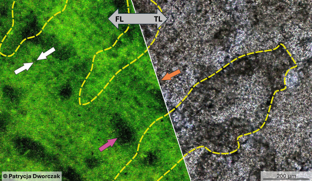
Modern LEDs and digital image processing in fluorescence microscopy make it possible to visualise skeletal structures that would otherwise be impossible (or very difficult) to detect in thin sections. Patrycja Dworczak, who is currently working as a postdoc with us at the GZN in Erlangen (Germany), has, together with her co-authors, applied this method to demonstrate that some Palaeozoic stromatoporoids originally possessed highly porous skeletons. The article is freely accessible and can be downloaded here:
https://link.springer.com/article/10.1007/s10347-026-00732-w
Palaeozoic stromatopores were much more porous than previously thought