Wear, tear and systematic repair: testing models of growth dynamics in conodonts with high-resolution imaging

Symbolic picture for the article. The link opens the image in a large view.
Reconstruction of growth dynamics obtained from a histological thin section of Ozarkodina confluens. (a) Morphological change calibrated with individual growth layers (lamellae of the crown tissue). (b) Composite BSE images of polished thin section (scale bar 50 µm) through the conodont element outlining three transects along which lamellae were counted. (c) Episodes of wear recorded as tip breakages. (d–g) Reconstruction of wear followed by repair of the damaged element. (h) BSE image of wear surface (scale bar 5 µm).

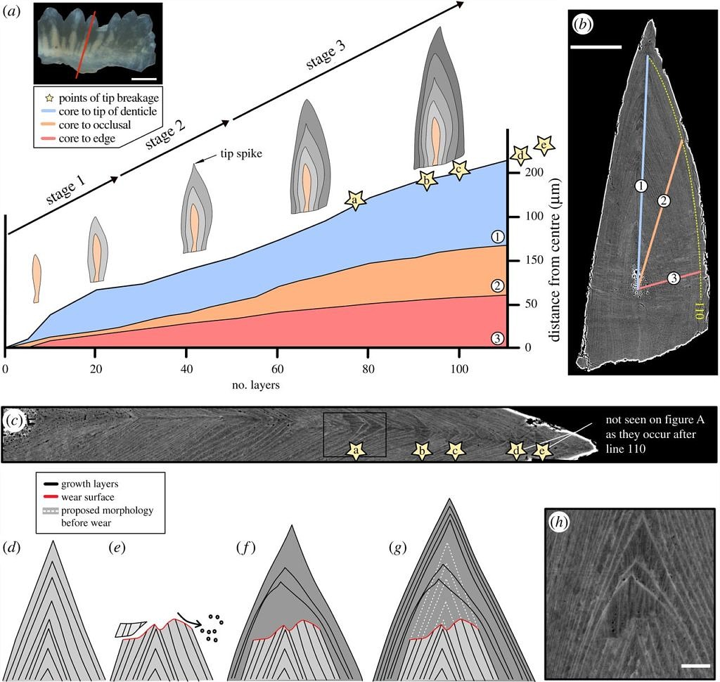
The first paper of our PhD student Bryan Shirley has been published in Proceedings of the Royal Society B. The co-authors are Madleen Grohganz, whose BSc thesis has contributed important data to the paper, Michel Bestmann from the Structural Geology group of the GeoZentrum, and Emilia Jarochowska. The paper employs innovative imaging methods to resolve a long-standing debate on the growth mechanism of conodonts and shows that these early vertebrates had a larval phase which was characterised by a different mode of feeding than the adult form.

Read the paper: Wear, tear and systematic repair: testing models of growth dynamics in conodonts with high-resolution imaging