Computer-Tomography-Laboratory
The Micro-Computer Tomography Laboratory at the Chair of Paleoenvironment
Geoscientific questions often require an examination of both the interior of rocks and the fossils they contain. Until now, traditional preparation methods have mainly involved the destruction of the sample material (sectioning, thin sectioning, and serial sectioning).
The emergence of computer tomography technology in the 1970s revolutionized medicine. Intensive research in this field since the 1980s has also led to the development of industrial computer tomography. Since the early years of the new millennium, this technology has become particularly interesting for sedimentological and paleontological/biological research, both in terms of its affordability and its performance. It is now possible to analyze and document the internal structures of rocks without destroying them.
Do you need scans of your samples for your research? Contact us: christian.schulbert@ fau.de oder telefonisch +49 9131 8524851.
Here are some examples from our laboratory that illustrate the possibilities offered by non-destructive material testing technology:
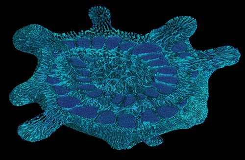
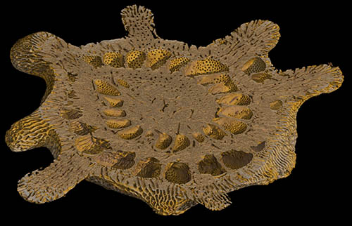
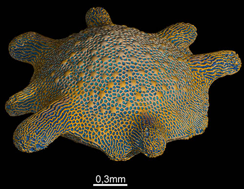
Morphological analyses of the shells of marine unicellular organisms in the submillimeter range. The raw 3D volumes are displayed in grayscale depending on the material density. Software can be used to segment this grayscale data into colors, which allows structures such as surfaces to be displayed more clearly. In the case of the recent foraminifer Calcarina, the calcareous shell and the cavity (blue) enclosed by the shell (yellow) are shown here.
Segmented volumes can also be shown and hidden, as well as virtually cut, to provide insights into the internal structures.
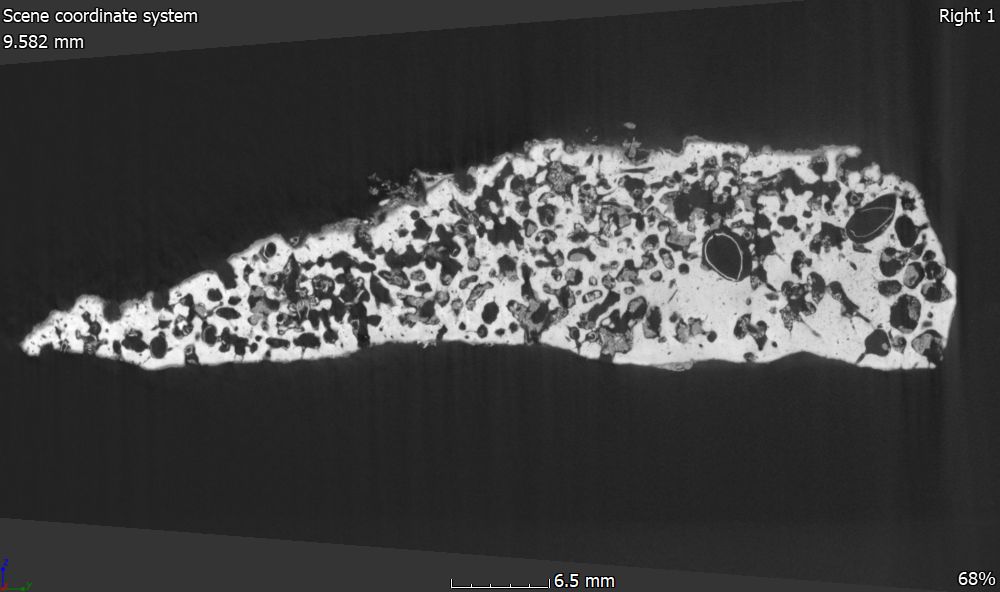
Bioerosion of organisms on other organisms or on inanimate substrates is widespread in both recent and fossil contexts. With the help of 3D volume data from CT scans, traces of bioerosion can be visualized and analyzed in a non-destructive manner. In this way, it was possible to identify the various groups that had hollowed out a limestone rock from the Mediterranean Sea. In the 2D section, for example, the shells of a boring mussel can still be seen in some cavities, which, along with many specimens of a boring sponge, caused the perforated structure of the once compact limestone.
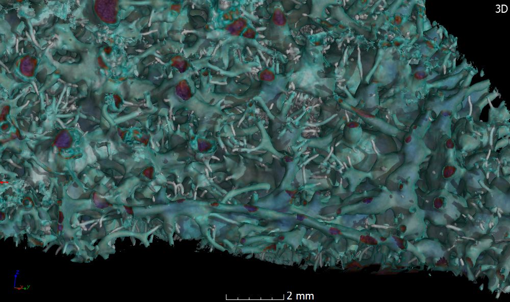
While a two-dimensional cross-section of an object already provides a wealth of information, this can be greatly expanded upon by using 3D representations of structures. Here, the cavities in the limestone have been vividly depicted, while the limestone itself has been removed or hidden. This structure was created by the marine boring sponge Cliona, which connects its many chambers with fine channels.
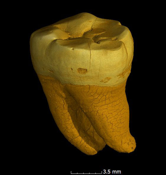
Our collection includes the wisdom tooth of the oldest human fossil found in Bavaria. Examinations of surrounding stalactites revealed an age of more than 250,000 years. At that time, representatives of our species were probably already living in the region, but Neanderthals were also widespread. Morphological measurements are necessary to determine which human species this tooth belongs to (the investigations are still ongoing).
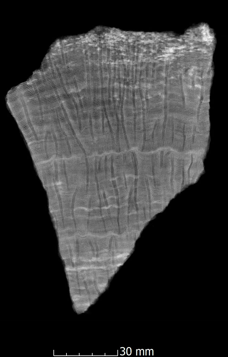
Coral skeletons are an excellent archive for climate research into temperature developments during the Pleistocene. Growth rates can be calculated, for example, using growth lines on the coral skeleton. In a seasonal rhythm, corals alternately build dense and less dense stripes into their skeletons. These differences in density can be easily detected and measured using computer tomography methods.
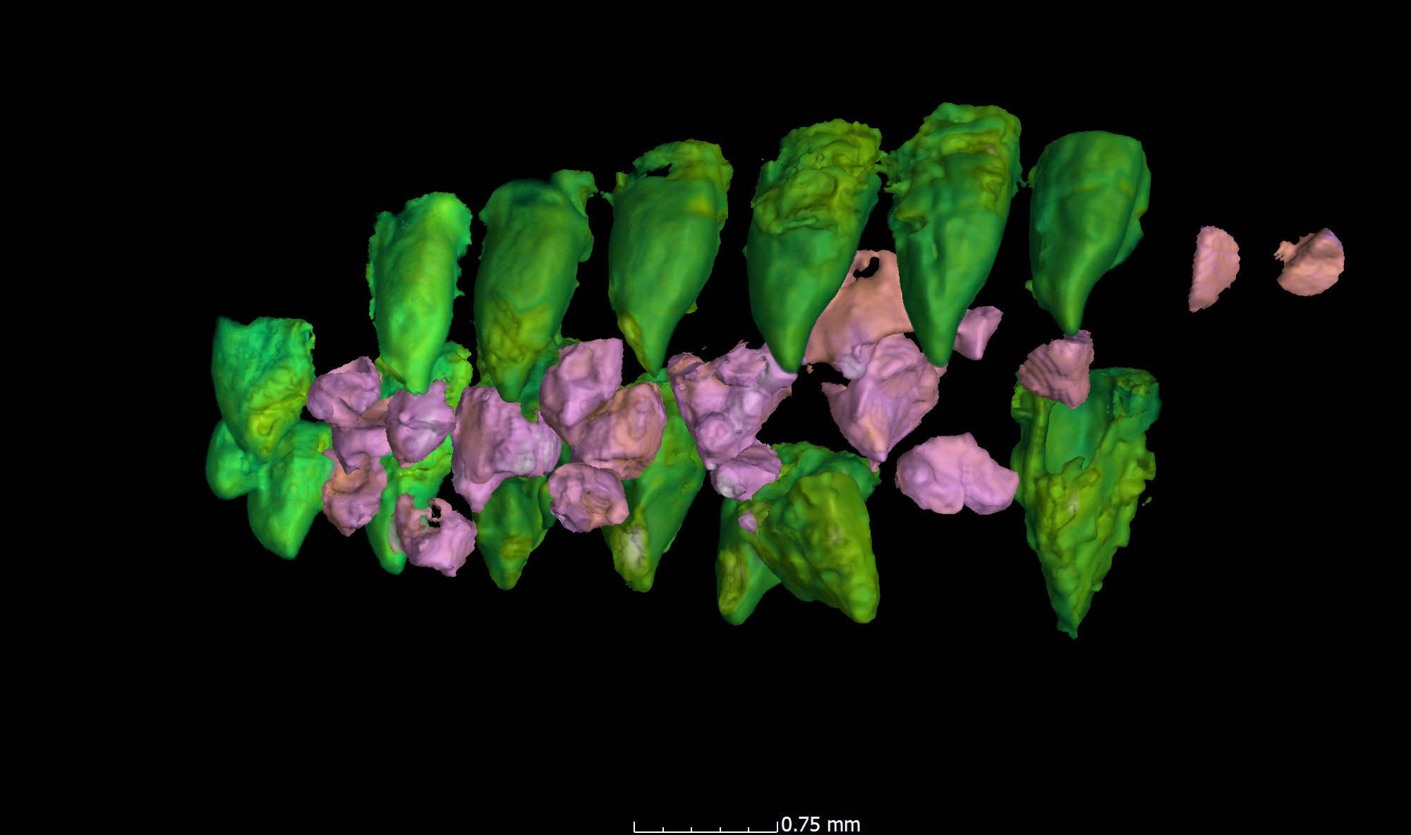
For vertebrate paleontologists, the internal structures of their objects of study are often inaccessible. However, in most cases, it is not possible to make thin sections and cuts through vertebrate fossils. In the case of the fish fossil Piranhamesodon from the limestone slabs of Ettling in the Altmühltal valley, it was only possible to determine that there were additional rows of teeth in its mouth on the palate using computer tomography methods. https://doi.org/10.1016/j.cub.2018.09.013
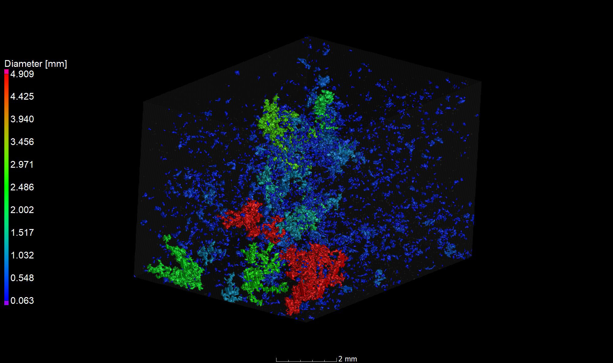
Sedimentologists can use CT data to visualize and measure the pore space in rocks. Pores and pore connections provide insights into the transport of liquids (or gases), such as groundwater or petroleum.
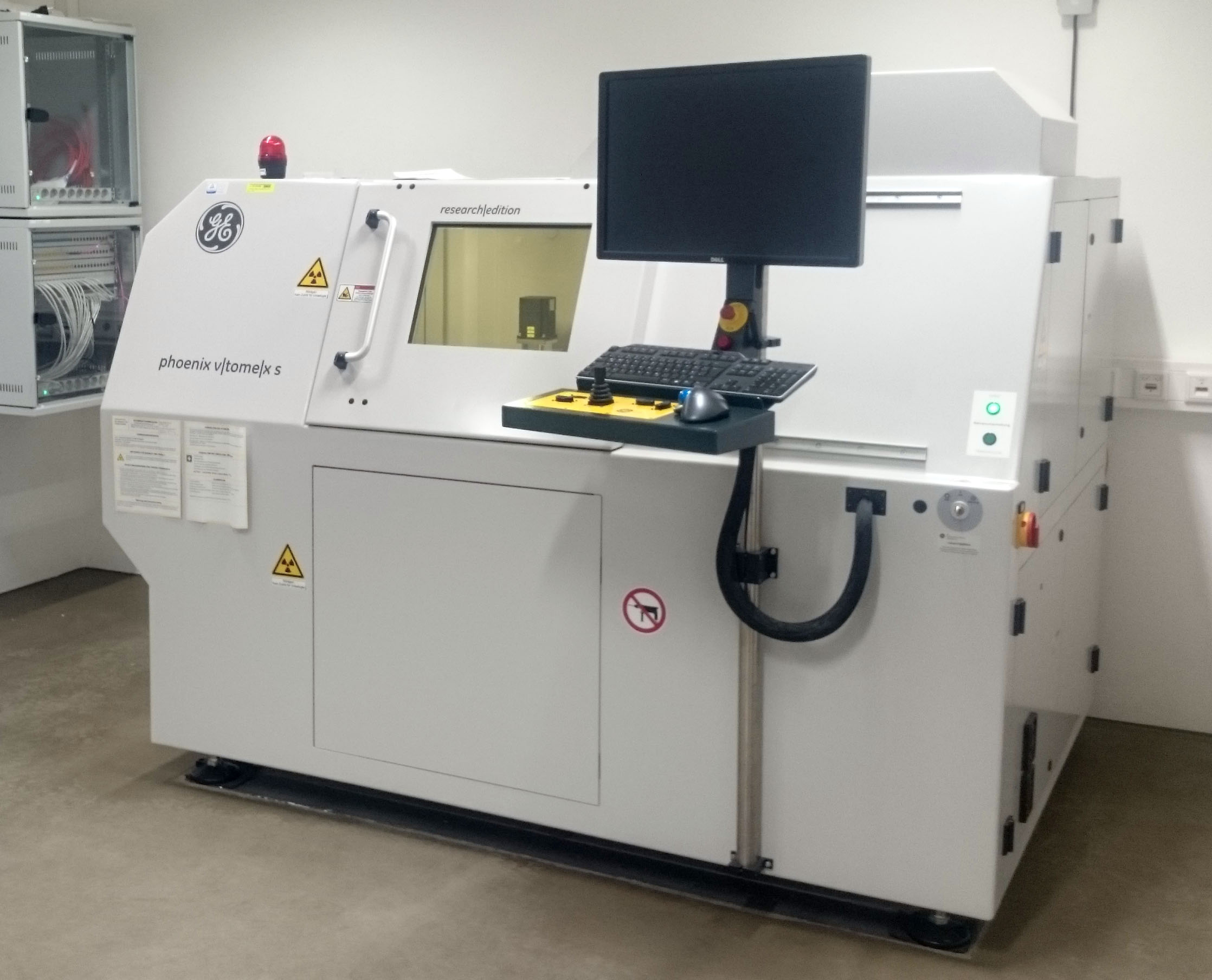
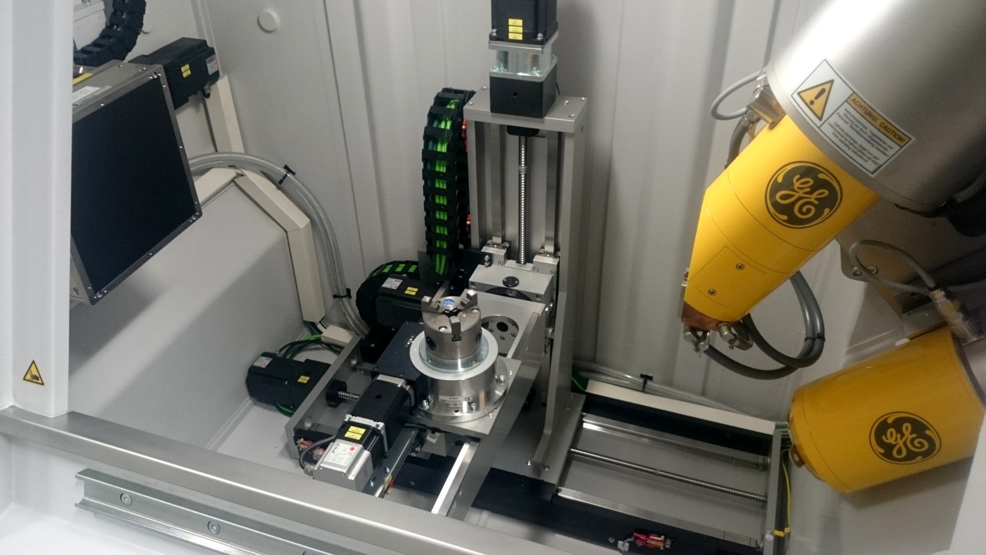
Micro computer tomograph
- Paläontologie
- Geologie
- Biologie
- Archäologie
- Medizin
- Mineralogie
- Materialwissenschaften
- Metallurgie
- Ingenieurswissenschaften
Model: Phoenix v|tome|x s 240
Construction Year: 2016
Location: Erlangen
URL: https://www.gzn.nat.fau.de/palaeontologie/ausstattung/computer-tomographie-labor/
Funding source: Deutsche Forschungsgemeinschaft (DFG)
Related Research Projects:
- Exploring the potential of coralline algae as climate proxy and for climate model evaluation: a Southern Hemisphere case study of New Zealand
- Size reductions during hyperthermal events: early warnings of environmental deterioration or signs of extinction? (EarlyWarn)
- FOR 2332: Temperature-related stresses as a unifying principle in ancient extinctions (TERSANE)
Related Publications:
- , , , :
The oldest deep-boring bivalves? Evidence from the Silurian of Gotland (Sweden)
In: Facies 65 (2019), Article No.: 26
ISSN: 0172-9179
DOI: 10.1007/s10347-019-0570-7 - , , , :
Microfacies analysis and 3D reconstruction of bioturbated sediments in the calcarenite di Gravina formation (southern Italy)
In: Marine and Petroleum Geology 125 (2021), Article No.: 104870
ISSN: 0264-8172
DOI: 10.1016/j.marpetgeo.2020.104870 - , , , , , , , , :
Instantiations of Multiscale Kinship in Pressing‐Defect Distributions in Yttria‐Stabilized Zirconias by Powder Partitioning
In: Advanced Engineering Materials (2024)
ISSN: 1438-1656
DOI: 10.1002/adem.202400139 - , :
Die moderne Computer-Tomographie (Modell v|tome|x s 240 von GE/Phoenix) am Lehrstuhl für Paläoumwelt (GZN, Erlangen) – Methodik und Anwendungsbeispiele
In: Geologische Blätter für Nordost-Bayern und angrenzende Gebiete 67 (2017), p. 249-258
ISSN: 0016-7797 - , , :
A new Pycnodontid (Actinopterygii) in the late Jurassic of the Solnhofen Archipelago
In: Research & Knowledge 3 (2017), p. 39-42
ISSN: 2408-204X - , , , , :
Coring tools have an effect on lithification and physical properties of marine carbonate sediments
In: Scientific Drilling 32 (2023), p. 43-54
ISSN: 1816-8957
DOI: 10.5194/sd-32-43-2023 - , , , , , , , , , :
Structural and geochemical assessment of the coralline alga Tethysphytum antarcticum from Terra Nova Bay, Ross Sea, Antarctica
In: Minerals 13 (2023)
ISSN: 2075-163X
DOI: 10.3390/min13020215 - , , , , , , , :
Evolution of the particle size distribution of tricalcium silicate during hydration by synchrotron X-ray nano-tomography
In: Cement and Concrete Research 156 (2022), Article No.: 106769
ISSN: 0008-8846
DOI: 10.1016/j.cemconres.2022.106769 - , , , :
Modern brackish bryostromatolites (“bryoliths”) from Zeeland (Netherlands)
In: Palaeobiodiversity and Palaeoenvironments (2021)
ISSN: 1867-1594
DOI: 10.1007/s12549-021-00490-3 - , , , , :
Morphological response accompanying size reduction of belemnites during an Early Jurassic hyperthermal event modulated by life history
In: Scientific Reports 11 (2021), Article No.: 14480
ISSN: 2045-2322
DOI: 10.1038/s41598-021-93850-0 - , , , , :
Biostratigraphy and sequence stratigraphy of the Toarcian Ludwigskanal section (Franconian Alb, Southern Germany)
In: Zitteliana 95 (2021), p. 57-94
ISSN: 0373-9627
DOI: 10.3897/zitteliana.95.56222 - , , , , , , , , , , , :
Decoding sea surface and paleoclimate conditions in the eastern Mediterranean over the Tortonian-Messinian Transition
In: Palaeogeography, Palaeoclimatology, Palaeoecology 534 (2019), Article No.: 109312
ISSN: 0031-0182
DOI: 10.1016/j.palaeo.2019.109312 - , , , , , , , , , , , , :
Following Changes at the Solid/Liquid Interface for Large Microplastic Particles by Streaming Potential
In: Chemistry Methods (2025), Article No.: e202500102
ISSN: 2628-9725
DOI: 10.1002/cmtd.202500102 - , , , , :
A new subdisarticulated machaeridian from the Middle Devonian of China: Insights into taphonomy and taxonomy using X-ray microtomography and 3D-analysis
In: Acta Palaeontologica Polonica 62 (2017), p. 237-247
ISSN: 0567-7920
DOI: 10.4202/app.00346.2017 - , , , , , :
The reefs of the Arctic - photoautotrophic ecosystem engineers endangered by microplastic and climate change?
The Arctic Biodiversity Congress (Rovaniemi, 9. October 2018 - 12. October 2018) - , , , , :
Growth interruptions in Arctic rhodoliths correspond to water depth and rhodolith morphology
In: Minerals 11 (2021), p. 1-13
ISSN: 2075-163X
DOI: 10.3390/min11050538 - , , :
3D-Analysis of a non-planispiral ammonoid from the Hunsruck Slate: natural or pathological variation?
In: PeerJ 5 (2017)
ISSN: 2167-8359
DOI: 10.7717/peerj.3526 - , , :
Rostrum size differences between Toarcian belemnite battlefields
In: FOSSIL RECORD 21 (2018), p. 171-182
ISSN: 2193-0066
DOI: 10.5194/fr-21-171-2018 - , , , , , :
Ultrastructure of the epidermal gland system of Tetranchyroderma suecicum Boaden, 1960 (Gastrotricha: Macrodasyida) indicates a defensive function of its exudate
In: Zoomorphology 138 (2019), p. 443-462
ISSN: 0720-213X
DOI: 10.1007/s00435-019-00462-4 - , , , :
Influence of different CA2/CA-ratios on hydration degree, AH3 content and flexural strength investigated for a binder formulation of calcium aluminate cement with calcite
In: Cement and Concrete Research 165 (2023), Article No.: 107090
ISSN: 0008-8846
DOI: 10.1016/j.cemconres.2023.107090 - , , , :
A Piranha-like pycnodontiform fish from the Late Jurassic
In: Current Biology 28 (2018), p. 1-6
ISSN: 0960-9822
DOI: 10.1016/j.cub.2018.09.013 - , , , , , , :
Microplastic pollution as a possible threat for an arctic reef system and its association of ecosystem engineers
54th European Marine Biology Symposium (Dublin, 25. August 2019 - 29. August 2019) - , , , , , , :
3D morphological analysis of cement over the first 24 hours of hydration by holographic and near-field ptychographic-tomography
ESRF Coherence Workshop (Grenoble, 9. September 2019 - 13. September 2019) - , , , :
Reef Fish Beware! Aggressive Mimicry in a Pycnodontid?
Annual Meeting of the Paläontologische Gesellschaft (München, 15. September 2019 - 18. October 2019)
In: Paläontologische Gesellschaft (ed.): Abstracts of the 90th Annual Meeting of the Paläontologische Gesellschaft, München: 2019 - , , , , :
What is boring? - Arctic reef structures as a habitat for boring organisms
The Arctic Biodiversity Congress (Rovaniemi, 9. October 2018 - 12. October 2018) - , , , :
Extreme reefs: Analyses of modern bryostromatolite ("bryolith") reefs from marginal environments in the Netherlands with comparisons to ancient analogues
EGU General Assembly Conference 2020 (Wien)
In: EGU General Assembly Conference Abstracts 2020
DOI: 10.5194/egusphere-egu2020-22617 - , , , , , :
Deeper insights: The potential of X-ray micro-computed tomography (μCT) in microplastic studies
MICRO 2020 - Fate and Impacts of Microplastics: Knowledge and Responsibilities (Lanzarote, 23. November 2020 - 27. December 2020) - , , , :
A rare find: protoconch in Cretaceous nerineid “gastropod dinosaurs”
90th Annual Meeting of the Paläontologische Gesellschaft (München, 15. September 2019 - 18. September 2019)
In: Paläontologische Gesellschaft (ed.): Abstracts of the 90th Annual Meeting of the Paläontologische Gesellschaft 2019 - , , :
Morphological characters and ontogenetic development of deep-sea species of the genus Caryophyllia
13th International Symposium on Fossil Cnidaria and Porifera 2019 (Modena, 3. September 2019 - 6. September 2019)
In: Università di Modena e Reggio Emilia, Dipartimento di Scienze Chimiche e Geologiche (ed.): 13th International Symposium onFossil Cnidaria and PoriferaModena, ABSTRACT BOOK 2019
URL: http://www.13thfossilcnidaria.unimore.it/wp-content/uploads/2019/09/Abstract-book-ISFCP-2019.pdf - :
Spurenlesen in der Vergangenheit - die Burghöhle Wolfsegg als Treffpunkt für Mensch, Höhlenlöwe und Co.
In: Mitteilungen - Verbandes der Deutschen Hohlen- und Karstforscher 69 (2023), p. 9-23
ISSN: 0505-2211